

Chlorophyll:
Collection and wet Lab:
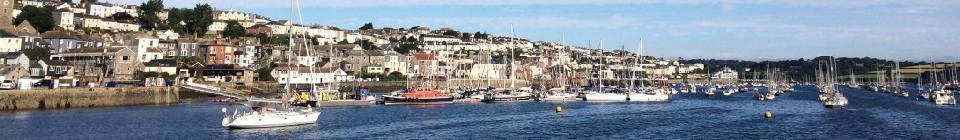
A 1000ml sample of seawater was collected from 1ft below surface using a Niskin bottle. A 60ml syringe was cleaned with 60ml of the respective seawater sample under testing and the water discarded overboard. 50ml of the seawater sample was then taken from the original sample bottle and a 0.2μm filter of 25mm diameter attached to the syringe to retain the organic matter from the extracted subsample. The filter paper was then removed, transferred and sealed in a test tube containing 90% acetone to form a lysate solution. This procedure was repeated once at each of the 4 stations on the river Fal at varying salinities (refer map) and these tubes were stored in a dark cool box for later analysis.
A shrimp larvae (centre) under a microscope

Lab analysis:
Chlorophyll samples taken from the previous day on the Winnie the Pooh using a horizontal
niskin bottle were placed into a glass tube and measured for fluorescence using a
10AU-
Phytoplankton:
Collection and wet Lab:
After triggering the water capture and returning the Niskin Bottle to deck containers used to store the water samples were rinsed to avoid contamination and transferred into glass bottles containing 1ml of Lugols for preservation and staining purposes (Williams et al., 2016) to be stored in a dark cool box to be transported to the dry lab.
Lab analysis:
To analyse these water samples for phytoplankton a 100ml sample was added to 1ml of Lugols for preservation and staining purposes (Williams et al., 2016). The top 90ml of the settled mixture was removed using a vacuum pump. The remaining 10ml of the solution was analysed, which theoretically contained all the phytoplankton cells from the 100ml sample.
A transfer pipette was used to place a 1ml into a Sedgwick Rafter cell, as a representative
volume of the 100ml sample. Each square in the cell contained a volume of 1μl. For
each sample, the phytoplankton contents of 100 out of the 1000 squares were identified
and recorded under a 10x magnification with a 10-
Coastal Plankton: Photo Guide for European Seas (Larink & Westheide, 2011) was used to ID the observed species on the cell. Multiplying the abundance of each species in the Sedgwick Rafter cell by 1000, created a value of abundance per litre.
Zooplankton:
Collection and wet Lab:
Direct samples of living zooplankton were collected using a plankton net (50cm diameter)
with a filtering size of 210μm for stations N, O, P and Q, and a mesh of 200 μm for
stations 33, 34, 35 and 36, which was lowered overboard by hand and towed behind
the vessel 1ft below surface for 5 minutes. After, the net was rinsed overboard with
seawater, filtered and retrieved. The sample was then fixed and preserved with 50ml
of 10% formaldehyde solution. This process was carried out 4 time, one in between
stations N and O, one between stations P and Q, both of which were towed counter-
Lab analysis:
To analyse the 1L water sample collected for zooplankton was inverted to mix up the settled cells to a homogenous mixture. A 10ml sample was removed using a 10ml measuring cylinder. From this sample, 2ml at a time was moved into a Bogorov Chamber using a 3ml transfer pipette to clearly identify and record the present phytoplankton species for the 10ml sample.
The chamber contents were viewed under a 10x magnification with a WILD M3Z light microscope (Serial No.: GY746). Coastal Phytoplankton: Photo Guide for Northern European Seas (Kraberg et al., 2011) was used to ID the observed species on the cell.
A calculation was completed for the species abundance to present the data as abundance
per m-
The volume of sea water sampled in m3 (V) is calculated by:
V = Π r2 L
Where r2 is the radius of plankton net opening (m), squared and L is the towing distance (m).
From this the number of zooplankton per m3 (N) is calculated by:
N = (n x 100)/ V
Where n is the number of zooplankton in 10ml of a 1L sample, V is the volume of seawater sampled (m3).


The results from the Niskin bottle water samples present that different phytoplankton
species dominate the water column at different positions throughout the Fal estuary,
during the same time of morning during the sampling day. Station Q is situated towards
the source of the Fal and is shown to be dominated by Mesodinium rubrum, at 65% (fig.
23). M. rubrum is a ciliate species that often causes red tides and is a significant
primary producer during bloom and and non-
At sampling Station 30, near the mouth of the Fal, M. rubrum composes a smaller percentage of the total phytoplankton population, of 12% (fig. 24). The dominant species at this station during the same sampling time is the diatom Nitzschia longissima (fig. 24). This station displays are more varied range of phytoplankton species, than the station towards the estuary source.
Figure 23. Percentage of phytoplankton species per litre of water sample collected at Station Q on 8th July at 09:24 (UTC), Location 50° 14.529' N 005° 01.549' W, water sample from a depth of 1.2m.
Figure 24. Percentage of phytoplankton species per litre of water sample collected at Station 30 on 8th July at 09:14 (UTC). Location at 50 08.688' N 005 01.416' W, water sampled from a depth of 1m.

Disclaimer: The views and opinions expressed are those of the contributors and do not reflect the views and opinions of the University of Southampton
Crawford, D, 1989, Mesodinium rubrum: the phytoplankton that wasn't, Marine Ecology
Progress Series, 58:161-
Williams, O, Beckett, R, & Maxwell, D, 2016, ‘Marine phytoplankton preservation with Lugol’s: a comparison of solutions’, Journal of Applied Phycology, 28, 1705.
Lanrink, O, Westheide, W, 2011, Coastal Plankton: Photo Guide for European Seas, 2nd, München: Pfeil Verlag.
Kraberg, A, Bauman, M, Dürselen, C-
Phytoplankton:
Zooplankton:

There is a difference in the recorded zooplankton abundance and species diversity at the two sampled stations of the Fal estuary. The figure (fig 25.) presents a high count of Copepods and Copepod nauplii for both Stations 33 and 36.
Note that the logged axis displays a large difference between the low abundances of 255 counts per m3 for Gastropod larvae, Siphonophorae, Ctenophora, Echinoderm larvae, Appendicularia and Fish larvae found in Station 36. However these species were not present in the samples further up the estuary, at Station 33, presenting that the zooplankton species are is more diverse towards the mouth of the estuary (see site map).
Figure 25. Logged zooplankton count per m3 of a 1m water sample, collected from the Fal Estuary on the 8th July. Station 33 sample collected at 11:52:54 (UTC), transect start location 50°12.873'N, 05°01.576'W. Station 36 sample collected at 14:01:27 (UTC), transect start location 50°08.788'N, 05°01.416'W.