Southerly winds and the forecast for the morning of the 26th of June 2015 gave us the opportunity to plan a trip almost 20 nautical miles south on board the RV Callista, farther than any groups had gone up to that point. However, in the morning the winds proved strong enough to create very rough seas with a large swell. Low tide was at 06:13 UTC, high tide was at 12:09 UTC.
The views and opinions expressed on this page are those of Group 3 and do not represent the views of the University of Southampton, the National Oceanography Centre or Falmouth Marine School.
The general trend for the Silicon profiles was for there to be lower concentrations in the surfaces layers and to increase with depth. There was an exception at station 34, where there was a decrease towards the mid depths and then an increase towards the seabed again. Station 34 decreased from 3.94 µmoles/l to 2.8 µmoles/l and then increased to 3.9 µmoles/l. This indicates an anomaly in the surface layer as it is much higher than the concentrations at other stations, this will most likely have been caused by a human error. Station 25 also has a decrease in concentration between the upper and middle samples, however it is much lower difference (0.06 µmoles/l). This is more likely to be caused by an actual inverted layer than by human error.
Diatoms and other organisms strip silicon at the surface for use in their frustules. At the bloom, there are increasing levels as although it is being utilised by organisms, on death they return silicon to the water column. At depth, light becomes a limiting factor in reproduction and growth thus less silicon is utilised. It may also be resuspended from sediments at depth.
Figures 1.1 & 1.2: Map and boat route (top), and a table of stations (bottom) (Google 2015)
The journey between station 25 (A) and station 26 caused significant seasickness issues with several group members. We therefore resorted to our plan B; to move back almost due north, coming into the shelter of the headland.
The plan from there was to take a transect back to our start point. This plan was
based on an unfortunate misunderstanding; the crew and the PI were under the impression
that our shakedown had occurred at the official reference point, Station B, where
all groups end their runs. This was not the case and this was realised in time to
set a new course for Station B. The end result is two variably detailed transects,
one moving north-
Unfortunately, due to a mix-


Figure 1.3 & 1.4: Images showing CTD deployment (top) and samples being taken from deployed niskin bottles (bottom).

Oxygen saturation should overall decrease with depth, this is due to the air-
Despite the surface ocean being subject to air interactions, oxygen levels are lower in the surface. This is due to respiration in the surface ocean utilising the oxygen. The highest levels tend to be at the depth at which our phytoplankton blooms were occurring as oxygen is being produced in large amounts by photosynthesis. At depth, the air sea interactions are at a minimum and no oxygen producing photosynthesis can occur (light limited). Therefore dissolved oxygen levels are low.
Turbidity was fairly consistent between stations at depths between 2 metres and 12 metres. However beyond this, there were large differences between stations, with station 29 displaying the highest turbidity at around 40 metres and 32 and 26 being the lowest at this depth. This spike may be due to the boats propellers increasing turbulence and bubbles.
Turbidity can be expected to be higher at depth due to the re-
Transmission corresponds with light attenuation through the water column. By 30m, almost all stations displayed a transmission of almost zero (station 32 showed a slightly higher value). Differences are slight, with station 32 and station 27 being the two extremes in our data, however the overall pattern is uniform between stations.
The high transmission in the surface waters is the euphotic zone. Below 10m, downwelling irradiance is significantly reduced, hence transmission is a lot lower than in the very surface layers. As depth increases, more wavelengths of the visible spectrum are absorbed and thus less and less continues to depth. By 10m, only one fifth of the light that entered is still in the visible spectrum, explaining the exponential decline in transmission across all our stations.
Fluorometry varied considerably with depth, as is to be expected as the values should be much greater where the phytoplankton bloom is occurring. Values were close to zero at the surface and at depth and increased to up to 0.75v at depths between 10m and 40m, depending on which station is considered. Station 25 saw the shallowest bloom, peaking at 20m and the strongest bloom was at station 34 (B) which occurred just over 30m. Station 34 also saw one of the most confined blooms, spanning only a small change in depth within the water column.
Phytoplankton blooms occur where nutrients and light are not limiting. At the very surface, nutrients are stripped from the water and thus are insufficient to support a large population of
Salinities across stations were very constant, with all salinities remaining within 0.6 PSU of one another. All stations seemed to show a slight increase in salinity with depth, due to the sinking of denser and more saline waters. Salinity spikes were evident at all stations but the most significant of these was at station 32. Data tended to be more uniform at the surface, with fewer salinity spikes present.
As with the temperature profiles, the depth of the mixing layer is very clear as this is the period in the shallows where salinities are fairly constant with few spikes characterising the plots. However, the mixed layer appears as 5m in our salinity data as opposed to a maximum of 10m shown on our temperature graphs. Salinity decreases with depth slightly as despite us being in the open ocean,
RV Callista
The nitrate concentrations followed a trend of low concentrations in the surface 20m of below 1 umol/L. At deeper depths the levels increase. At stations 25 and 28 the values for the deepest samples are particularly high at over 3 umol/L. Station 30 is the only station that does no follow the general increase in concentration with its peak occuring at mid depth of around 30m before returning to low levels again at depth.
High levels of phytoplankton in the upper layers strip essential nutrients from the surface, as with phosphate. The conditions in the upper layer are ideal for phytoplankton growth so large blooms will strip the water of nitrate. At depth the conditions are less ideal for phytoplankton growth so less nitrate is taken out of the water, hence the increasing concentrations as depth increases (Garside,1985). Station 25 is the closest station to land therefore its high levels at depth may in part be due to anthropogenic sources.
The phosphate concentration between stations followed a similar gradient; lower at the surface and then increasing with depth. This makes sense as the phosphate would be rapidly scavenged by phytoplankton at the surface but less as light levels diminish. Only station 28 does not follow this pattern, having a higher value at the surface than at depth, almost certainly indicating an outlier.
Phosphate is scavenged in the surface by phytoplankton, therefore levels are extremely low. These organisms are ‘survivalists’ as afore mentioned in the fluorometry discussion for open ocean (Arrigo, 2005). At depth, phosphate levels are the greatest where light levels are limiting and phosphate is therefore not stripped from the water column.


Acoustic Doppler Current Profiler (ADCP)
ADCP transects were conducted between stations using an ADCP mounted on the hull
of RV Callista. The transects taken were then examined within specialised ADCP WinRiver
computer software to locate the position of the front. The front exists at the breakdown
of the thermocline and a well-
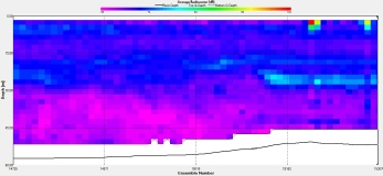
The ADCP backscatter shown in figure (2.6) increases at depth near the seabed, which is likely the result of an increased amount of sediment resuspension from the seabed. This could be caused by changes in seabed topography as the water depth increases between stations 25 and 26. Figures (2.9 and 2.12) show concentrated regions of intense backscatter at the surface, which could be flow noise due to cavitation caused through directional changes of RV Callista while steaming (Heywood et al, 1991). The large swell and high wind speeds encountered during this day offshore will have likely produced air bubbles in the surface waters that could have contributed to high levels of backscatter at the surface. The ADCP transects undertaken show no clear evidence that we crossed over the front, as there does not appear to be intense subsurface backscatter which would imply the presence of a phytoplankton and subsequent zooplankton blooms.

Figure 2.6: Backscatter of ADCP transect between stations 25 and 26.

Figure 2.7: Backscatter of ADCP transect between stations 26 and 27.

Figure 2.8: Backscatter of ADCP transect between stations 27 and 28.

Figure 2.9: Backscatter of ADCP transect between stations 28 and 29.


Figure 2.10: Backscatter of ADCP transect between stations 29 and 30.
Figure 2.11: Backscatter of ADCP transect between stations 30 and 31.


Figure 2.12: Backscatter of ADCP transect between stations 31 and 32.
Figure 2.13: Backscatter of ADCP transect between stations 32 and 33.

Figure 2.14: Backscatter of ADCP transect between stations 33 and 34.

Temperature was fairly consistent across the stations, ranging from a surface temperature of 15.5°C to 14.6°C, apart from at station 27. Station 27 saw a surface temperature of less than 14°C, marking a very clear front. All stations clearly show the thermocline and temperatures suddenly decrease on reaching it. The depth of the thermocline varies from around 6m at station 25 to just over 10m at most other stations. Station 26 saw the warmest temperature at depth at 12.6°C.
In the surface 10m, mixing has a strong influence shown by the uniform temperatures above the thermocline. Mixing occurs most significantly in these surface layers due to the interaction between
the water and the overlying air. The friction between the two mediums causes turbulent mixing of the surface layers, the influence of which decreases further from the interface (Brainerd, 1995). Temperature becomes much less uniform with depth at the thermocline. Uniformity in temperature returns on reaching the deep water temperature. Whilst the deep water temperature varies between our stations, the depth at which the temperature becomes constant again is at approximately 30m across all stations.
there still may be a small amount of influence from the nearby estuary inputting fresher waters onto the surface. The salinity spikes characterising the data are caused by a difference in sensor response times for temperature and salinity on the CTD (AML Oceanographic, 2015). Larger spikes indicate a larger difference between sensors.
plankton. At our stations, this nutrient limiting layer spans from a depth of 0 to 20m. Fluorometry is not at zero as some species can cope with low nutrient levels due to possessing large quantities of resource acquisition machinery (the ‘survivalist’) (Arrigo, 2005). Below the bloom light is the limiting factor, inhibiting photosynthesis and thus preventing large phytoplankton populations. Between 20 and 40m was where most of our stations saw the bloom. This is the region where neither light nor nutrients are limiting growth and reproduction.
- There is a very large species diversity in the structure of phytoplankton communities, both in terms of location and depth.
- The most abundant zooplankton group overall was the Copepoda, but at two stations (27 and 34) the Cladoscera were the dominant group. This is unusual, and could be attributed to human error while counting.
- Among the tested nutrients (silicon, phosphate, and nitrogen), most followed the general pattern of low concentrations at the surface, with the concentration increasing with depth. This could be because of biological uptake at the surface and remineralisation at depth.
- There is a large amount of variability between stations with regard to oxygen saturation.
- A mixed surface layer is shown at each station, with uniform temperature lying above the seasonal thermocline and station 27 displaying the deepest mixed layer.
- Salinity appears to be fairly similar between each station, each increasing slightly with depth.
- All stations show a deep chlorophyll maximum between approximately 25 and 35m depth.
- Transmission is highest at the surface, before decreasing with depth.
- Turbidity decreases with depth at all stations.


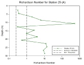
The Richardson number (Ri) is a dimensionless number which describes the ratio between a buoyancy force term and a flow gradient term (the equation for which is shown in figure 2.15). It can be used to estimate stability and acts as a guide to determine where turbulent mixing is likely within the water column and where thermal stratification may prevent mixing from occurring. Where Richardson numbers are small (<0.25) mixing is likely to take place, while larger values (>1) indicate where vertical mixing is unlikely and laminar flows will likely dominate. Between these two values is a transition zone. On each of the graphs these thresholds are indicated to make distinction clearer. The Richardson number values were calculated using flow velocity data acquired by the ADCP and density data collected by the CTD.
Comparing the Richardson number profiles from Stations 25-
Figure 2.1: Temperature depth profiel
Figure 2.2: Salinity depth profile
Figure 2.3: Fluorometry depth profile
Figure 2.4: Transmission depth profile
Figure 2.5: Turbidity depth profile
Figure 2.15: Station 25 (A)
Figure 2.16: Station 26
Figure 2.17: Station 27
Figure 2.18: Station 28
Figure 2.19: Station 29
Figure 2.20: Station 30
Figure 2.21: Station 31
Figure 2.22: Station 32
Figure 2.23: Station 33
Figure 2.24: Station 34 (B)
Figure 3.1: Silicon depth profile
Figure 3.2: Phosphate depth profile
Figure 3.3: Nitrate depth profile
Figure 3.4: Oxygen depth profile


Figure 2.15: Equation to calculate the Richardson number
Buoyancy force term
Flow gradient term
Phytoplankton
The phytoplankton samples taken from the Callista show a huge amount of species diversity, both in location and depth. For example, Nitzschia longissima is very dominant at stations 27, 28, and 34, but only present as a minority at stations 25 and 30. Along with this, at station 28, Nitzschia sp. is only present at 31.4m, and not present at 10.3m or 49.4m. As the three samples were taken to be representative, with one above, below and on the subsurface chlorophyll maximum, the fact that Nitzschia sp. only occurs at the middle depth shows that this species was making up the majority of that bloom.


Figure 3.5: Graphs showing phytoplankton results for stations 25, 27, 28, 30 and 34
Over all 5 stations by far the most abundant group was the copepoda, sometimes numbering upwards of 6000 individuals per square metre. It should be noted however, that the copepod count was a combination of two categories; the copepoda and the copepoda nauplii which is a different life stage of the same group. This is because while counting some members of the group counted the nauplii as a different category while others did not.
Station 27 and Station 34 are interesting as they show high numbers of cladocera in conjunction with high numbers of copepoda, relative to the relevant stations. The cladocera outnumber the copepoda at both stations. This is unusual as the cladocera are in low numbers or absent from other stations. This could indicate some form of human error, perhaps in the misidentification of other groups as cladocera. This may have resulted in higher or lower counts.


Figure 3.6: Graphs showing zooplankton results for stations 25, 27, 28, 30 and 34
References
Heywood, K.J., Scrope-
Brainerd, K., Gregg, M. (1995) Surace mixed and mixing layer depths. Deep Sea Research
Part I: Oceanographic Research Papers. Vol. 42(9), pp: 1521-
AML Oceanographic. (2015). Response times and calculated parameters. Website. http://www.amloceanographic.com/Technical-
Arrigo, K.R. (2005). Marine microogranisms and global nutrient cycles. Nature. Vol.
437. Pp: 349-