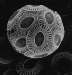
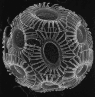
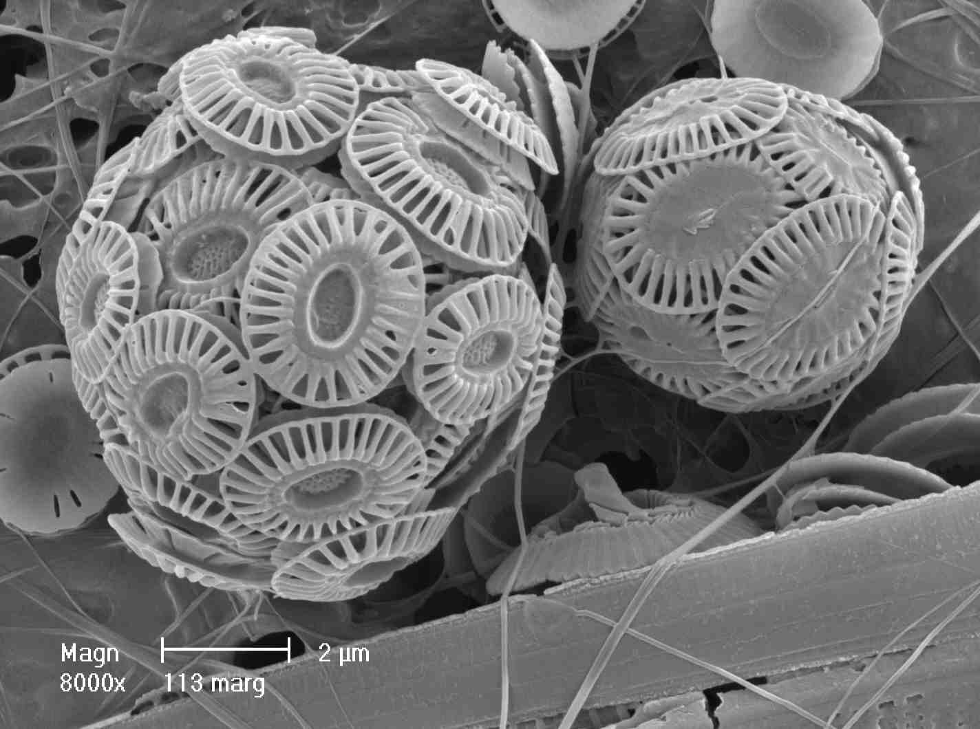
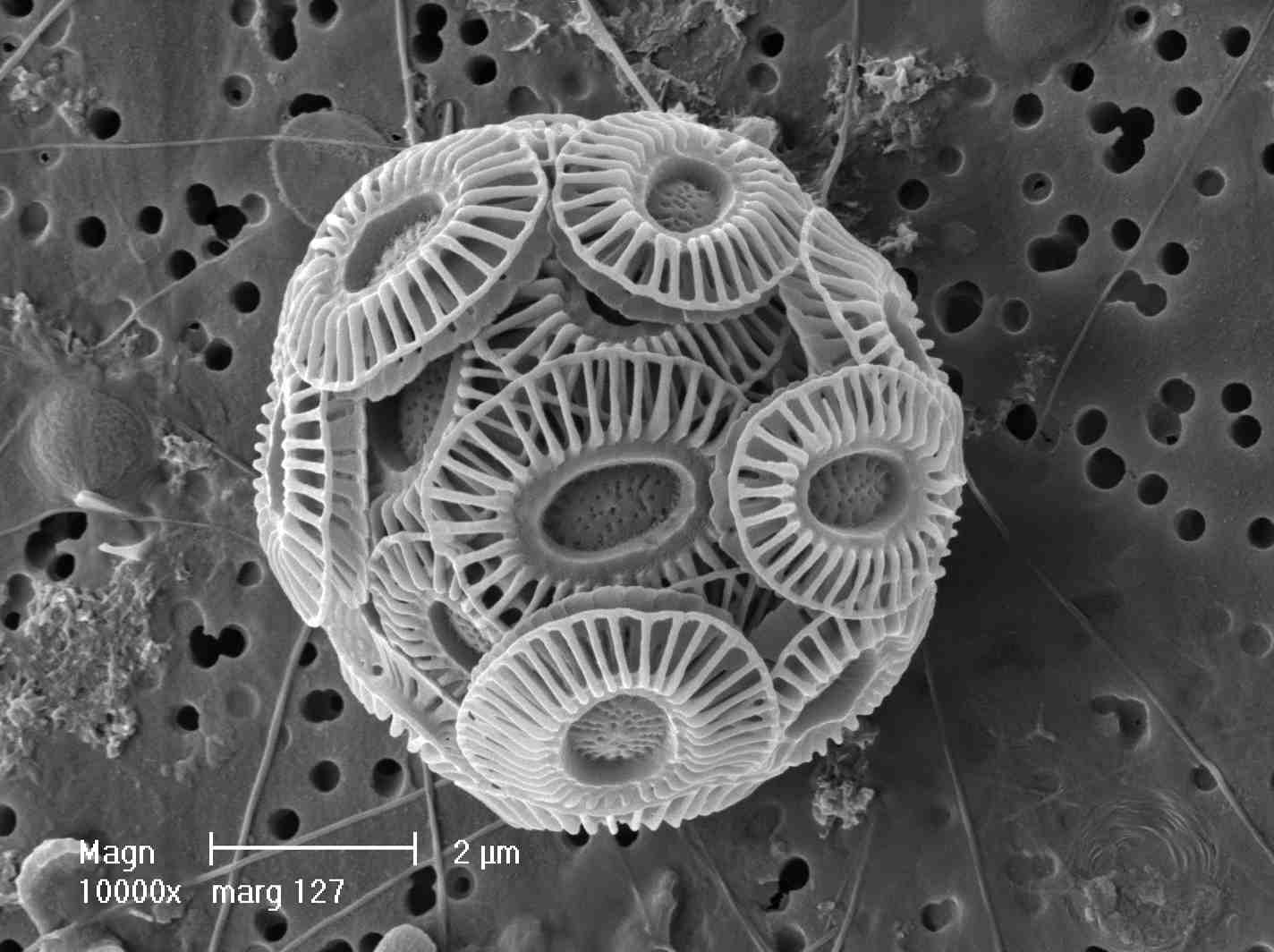
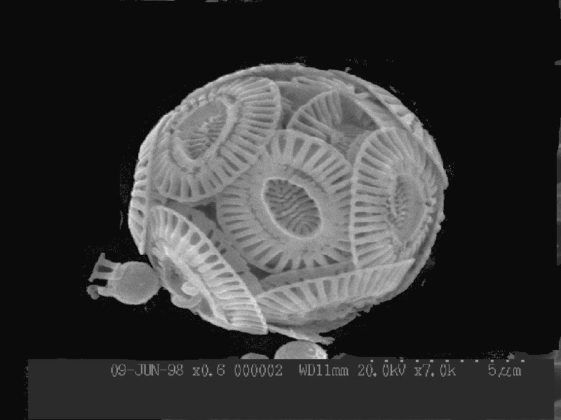
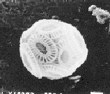
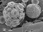
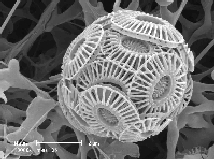
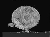
SEM pictures of Emiliania huxleyi Cells
Some of the pictures below show very tight, compact, cohesive coccospheres,
whereas some of the others are much looser, with coccoliths apparently
breaking away. Variation between coccospheres may be due to age of the
cells, different growing conditions, to genetic differences between
different strains of Ehux, or even to partial dissolution
(dissolving) of the calcite. The last coccosphere is known to be from a
different genetic strain than some of the others.
[click on the small pictures on this page to view them at full size]
The small white bar at the bottom-right of some pictures is a scale bar of
length 1 micron (1 x 10⁻⁶ metres).
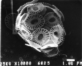
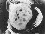


(top three pictures from Patrick Holligan, next five from Jeremy Young, last
from Luis Lampert).
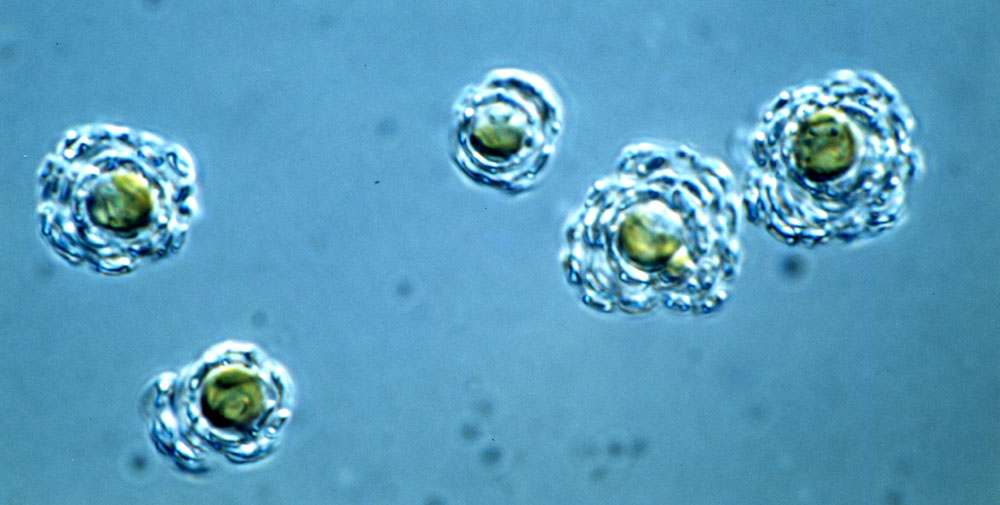
For comparison below is a light microscope picture of some calcified
(coccolith-covered) Ehux cells. Through most light microscopes Ehux appears
as nothing more than a fuzzy pin-head, even at highest magnification. This
is as good as it gets, from a state-of-the-art Leitz Orthoplan using
Differential Interference Contrast (DIC). The coccoliths are fairly but not
completely transparent (to light as opposed to electrons), and in this case
multiple layers of coccoliths are doubling or tripling the overall cell
diameter. The cells are a typical prymnesiophyte golden or golden-brown
colour (Photo from Ian Probert).

Ehux
home page
Toby Tyrrell : T.Tyrrell@noc.soton.ac.uk