Description of an Emiliania huxleyi cell
Jeremy Young
Palaeontology Dept.
The Natural History Museum
London, SW7 5BD
Great Britain.
Email: j.young@nhm.ac.uk
[click on the picture to see it full size]
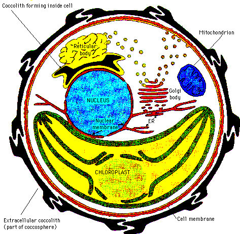
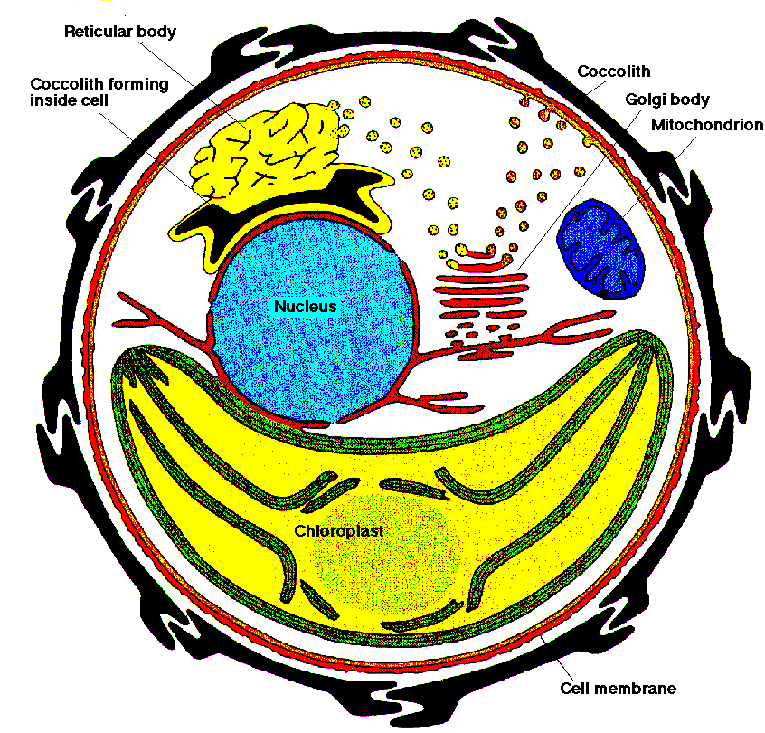
Schematic of Ehux cell: ER = endoplasmic reticulatum; (diagram from Peter
Westbroek)
Organelles: this drawing shows a cross-section through a single
Emiliania huxleyi. It is based mainly on observations made during
transmission electron microscope studies of ultrathin (/<1 micron) sections
of cells. As the figure clearly shows Ehux is unicellular, i.e.
formed of a single cell, but this cell contains a number of internal
organelles. Dominating the cell is the chloroplast - the photosynthetic
body. This contains photosynthetic pigments and works in
essentially the same way as chloroplasts in land plants, although the
pigments are slightly diferent so the colour is yellow or golden brown
colour rather than green. In the centre of the cell, above the chloroplast
is the nucleus. This contains the genetic material, DNA and is the site
for replication of DNA during cell division and production of RNA. The
mitochondrion is a distinct organelle, present in all eukaryotic cells,
and is the source of ATP (adenosine triphosphate) the major energy
transferring molecule of biological systems.
Membranes: the nuclear membrane connects with an internal
network of membranes, the endoplasmic reticulum [ER] and through this to the
cell membrane. All these are formed by the Golgi Body. By
contrast the chloroplast and mitochodrion have discrete embranes which
separate them from the rest of the cell, and they contain separate DNA.
These indicate that the chloroplasts and mitochondrion represent once
discrete singe-celled organisms which were incorporated via endosymbiosis
into the host protist. These endosymbioses are now thought, at least in the
case of coccolithophorids, to have occurred only once, in the distant
evolutionary past of the group's ancestors.
Coccolith formation: the most distinctive feature of the cell is the
covering of coccoliths. The coccoliths are formed inside the cell via a
highly organised process, coccolithogenesis. As shown, only one coccolith
at a time is formed in Ehux and they develop within a coccolith
vesicle close to the nuclear membrane. Above the coccolith vesicle is the
Reticular Body which controls the coccolith formation. This is quite
separate from the Golgi Body but is closely related to it, and microvesicles
can be observed between the two organelles. In other coccolithophorids the
coccolith vesicles form inside the golgi body without a distinct reticular
body being present. When the coccolith is fully formed the coccolith
vesicle migrates toward the edge of the cell, the vesicle mebranes fuse with
the cell membrane and the external parts of the membrane are resorbed so
that the coccolith is extruded to the outside of the cell.
Further Reading: for more information, see (Green & Leadbeater, 1994;
Green et al, 1989; Jordan et al, 1995; Pienaar, 1994; and refs in Westbroek,
1984).
References
Ehux
home page